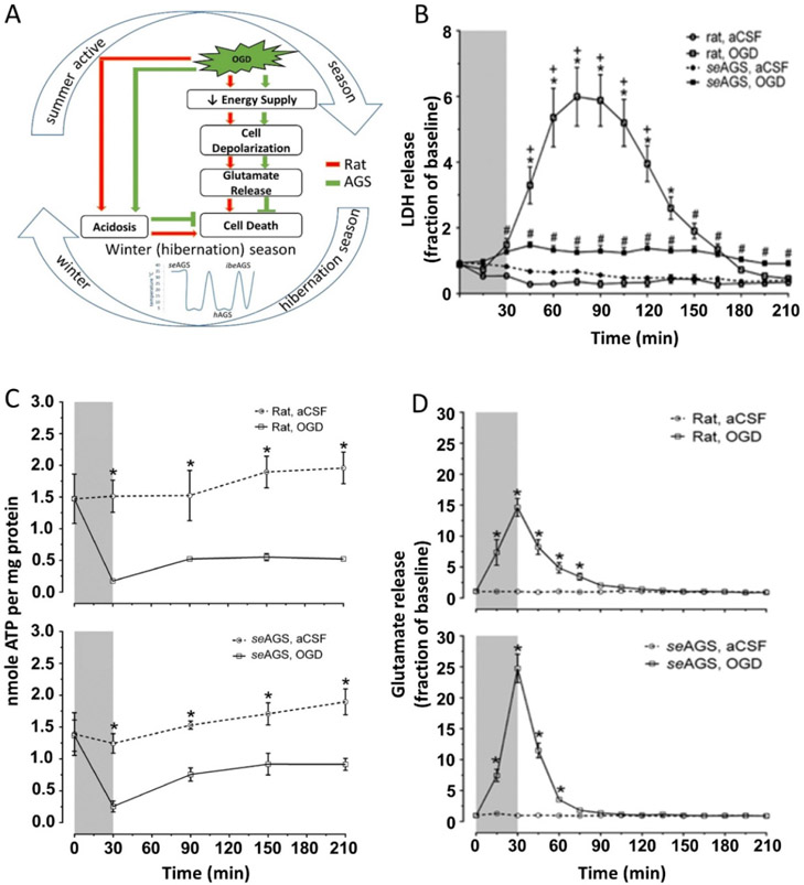
Figure 1.
AGS tolerate OGD injury despite ATP loss and glutamate efflux (A) Schematic representation of possible pathway in AGS tolerance to injury. (B) Lactic acid dehydrogenase (LDH) in perfusates collected every 15 min increases in rat hippocampal slices exposed to oxygen-glucose deprivation (OGD) (rat, OGD), but not in rat slices exposed to artificial cerebral spinal fluid (aCSF) (rat, aCSF), nor in slices harvested from summer euthermic AGS and exposed to aCSF (seAGS, aCSF). A small amount of LDH is released from slices collected from seAGS and exposed to OGD (seAGS, OGD). *p < 0.05 rat aCSF vs. rat OGD, +p < 0.05 rat OGD vs. seAGS OGD, #p < 0.05 seAGS aCSF vs. seAGS OGD. (C) Levels of whole tissue ATP were determined over time following bath application of treatment in slices from (a) rat (aCSF versus OGD) and (b) seAGS (aCSF versus OGD) subjected to either 30 min of aCSF or OGD followed by 3 h reperfusion. Gray bar indicates insult period. Data shown are means ± SEM, *p < 0.05 versus aCSF. (D) Time-dependent excitatory neurotransmitter (glutamate) efflux in rat and seAGS hippocampal slices induced by 30 min OGD insult. (n = 17 slices from six rats, n = 14 slices from five seAGS). Gray bar indicates insult period. *p < 0.05 for OGD versus aCSF group. Data shown are means ± SEM. [Reprinted with permission from Bhowmick et al. 2017a.]

