
Fig. 4.
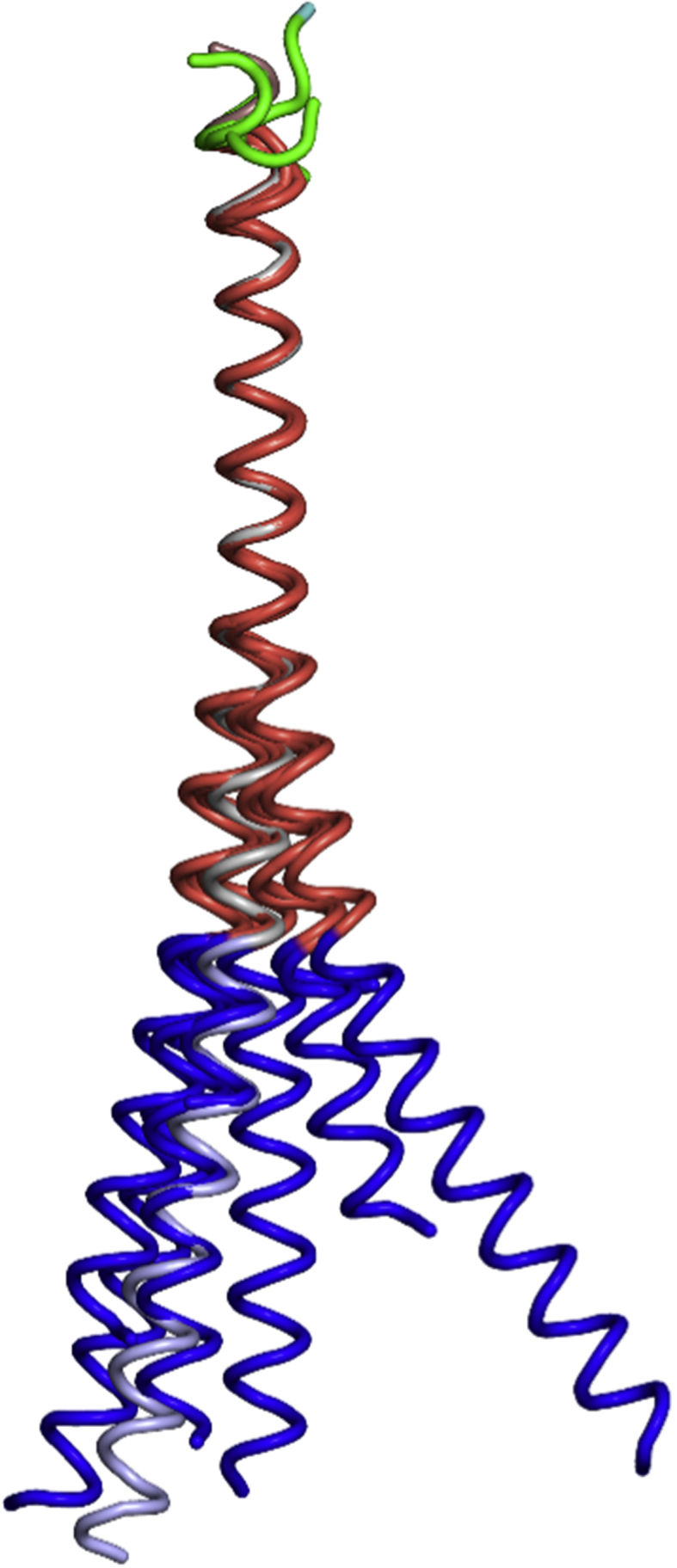
Superposition of FB subunits reveals conformational variability. Superposition of all eight independent FB monomers, FBSS (A, B, C, D), FB Type I (A) and FB Type II (A, B, C) using K194 – Arg210 (which contains H3 – H4). In grey, FB from the ΔFosB/JunD bZIP + DNA structure is also shown (PDB ID: 5VPE).