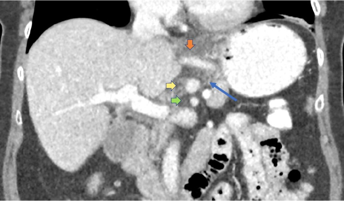
Figure 1.

Initial CT abdomen and pelvis with intravenous contrast performed in standard portal venous phase (70–80 s postcontrast injection); coronal two-dimensional reformatted image through upper abdomen. Abnormal perivascular hazy fat stranding (long blue arrow) seen as increased soft tissue attenuation around the mesenteric arteries. This is a non-specific finding that can reflect oedema, inflammation or cellular infiltration. Limited detailed assessment of the arterial walls due to venous timing of imaging. Orange vertical arrow: left gastric artery and accessory left hepatic artery. Yellow horizontal arrow: coeliac artery. Green horizontal arrow: superior mesenteric artery.
