. 2020 Jun 2;9:e53913. doi: 10.7554/eLife.53913

Figure 3. TMEM95 does not interact with JUNO nor IZUMO1.

(A) Binding analysis using the AVEXIS assays shows that the soluble recombinant TMEM95 ectodomain does not interact with JUNO nor with IZUMO1. The entire ectodomains of the named proteins were expressed in HEK293-6E cells either as biotinylated baits or as pentameric beta-lactamase-tagged preys. Bait proteins were immobilised on streptavidin-coated plates and captured prey proteins quantified by measuring the absorbance of a colorimetric reaction product of the beta-lactamase substrate, nitrocefin. The CD200R (bait)-CD200 (prey) binding pair was used as positive control. The same prey, CD200R, was tested against TMEM95 and is shown as negative control. Bars represent means + s.d.; n = 3. (B) HEK293 cells stably expressing the N-terminal half of GFP (GFP1-7) and mouse JUNO stained with a highly avid IZUMO1 probe. (C) HEK293 cells stably expressing the C-terminal half of GFP (GFP8-11) and mouse IZUMO1 stained with a highly avid JUNO. (D) TMEM95 does not induce fusion when expressed in HEK293T cells in the presence of JUNO and IZUMO1 using a GFP-complementation cell fusion assay. HEK293T cells expressing either half of GFP and either JUNO or IZUMO1 were mixed and their fusogenic ability visualized by GFP fluorescence. The IZUMOI-expressing cells were either mock transfected prior to mixing (Control), transfected with Syncitin a, as a positive fusion control, or Tmem95. By contrast to the cells transfected with Syncytin a, Tmem95 did not induce cell fusion. Cell nuclei are stained with DAPI and scale bar represents 20 µm.

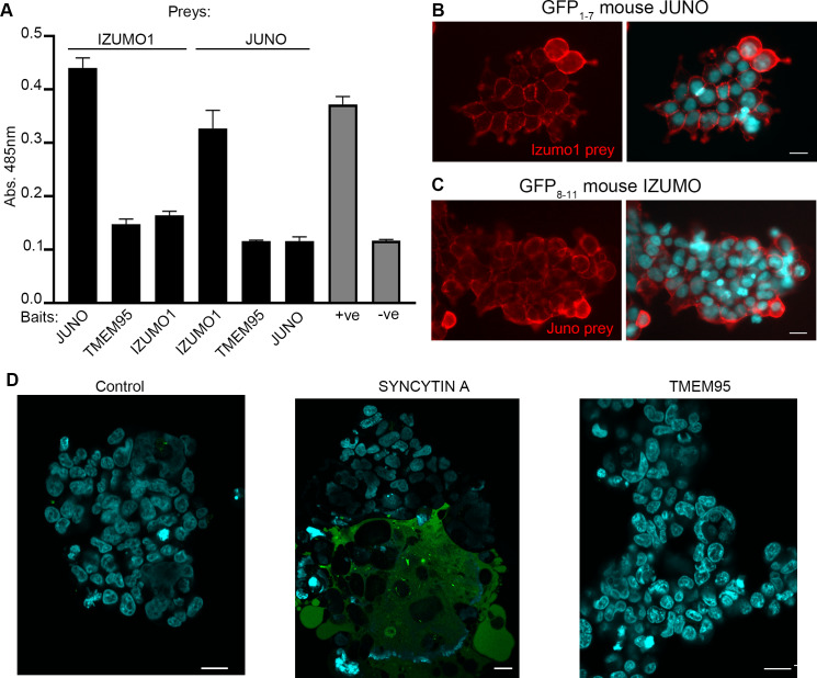
Figure 3.

Figure 3—figure supplement 1. Split GFP fusion assay.

Figure 3—figure supplement 1.

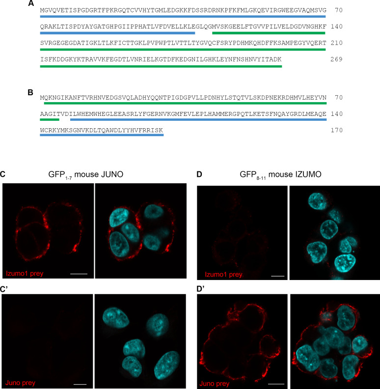
(A) Amino acid sequence of FKBP1A underlined in blue fused via a short linker to the N-terminal region of GFP (GFP1-7) (green underline). (B) Amino acid sequence of the C-terminal region of GFP (GFP8-11) highlighted in green, fused to RB, underlined in blue. (C, C´, D and D´) The cells expressing the GFP1-7 stably express mouse Juno and the cells expressing the GFP8-11 stably express mouse Izumo1. To establish functional cell surface expression of both Juno and Izumo1 on the cells, noth cell lines were stained with the corresponding pentameric FLAG-tagged Izumo1 and Juno preys. The cells expressing mouse Juno were stained with the Izumo1 prey but not Juno, and the cells expressing mouse Izumo1 stained with the Juno prey but not Izumo1. Images are representative of at least three independent replicates. Nuclei are stained with DAPI (blue) and scale bars represents 10 µm.
Figure 3—figure supplement 2. Sequence alignments of TMEM95 and IZUMO1.

Figure 3—figure supplement 2.

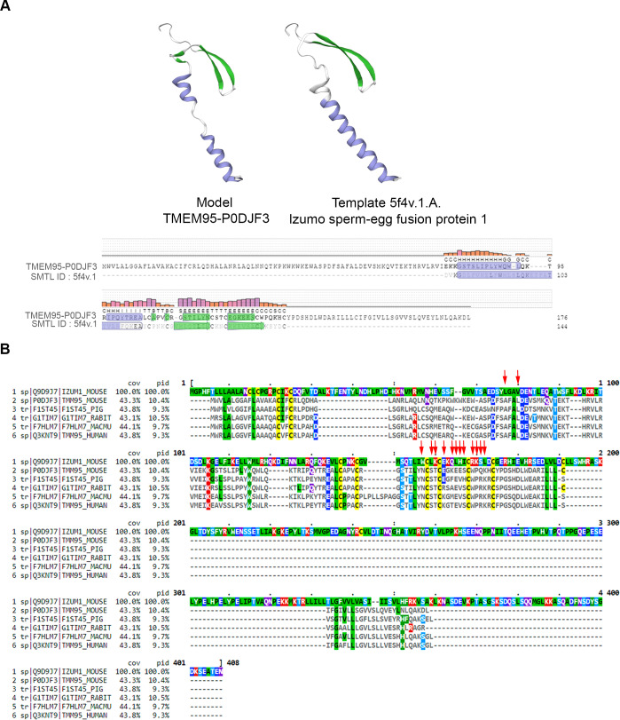
(A) Structure prediction of TMEM95 created by SWISS-MODEL web-based integrated service. Model TMEM95-P0DJF3 (left) was built using Template 5f4v.1.A (right) from Izumo1 sperm-egg fusion protein 1 (Ohto et al., 2016). Secondary structure alignment shows coloured residues by secondary structure elements: green for β-hairpin and blue for α-helix. (B) Sequence alignment of IZUMO1 from Mus musculus (Q9D9J7) (top) and TMEM95 from Mus musculus (P0DJF3), Sus scrofa (F1ST45), Oryctolagus cuniculus (G1TIM7), Macaca mulatta (F7HLM7) and Homo sapiens (Q3KNT9) are shown. Residues are colored by identity to top sequence (IZUMO1) and to indicate physicochemical properties: dark-gray for mismatch, yellow for cysteine, green for hydrophobic, dull-blue for small alcohol, bright-blue for negative charge, red for positive charge, and purple for polar. The residues of the IZUMO1-JUNO interface binding (Ohto et al., 2016) are indicated by red arrows. cov, percentage of coverage; pid, percentage of identity with top sequence. The sequence identity between mouse and human TMEM95 is 67%.