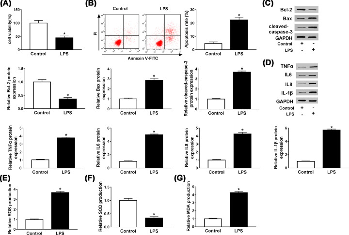
Figure 1. LPS reduced cell viability and induced apoptosis, inflammatory injury and oxidative stress in HUVECs.
(A,B) MTT and flow cytometry assays were used to determine the viability and apoptosis of LPS-induced HUVECs, respectively. (C,D) The levels of apoptosis-related proteins Bcl-2, Bax, cleaved-caspase3 and inflammation-related cytokines TNFα, IL-6, IL-8 and IL-1β in LPS-treated HUVECs were detected by western blot. (E–G) The productions of ROS, SOD and MDA were measured by DCFH-DA method and a commercial kit. *P<0.05.

