Figure 2.
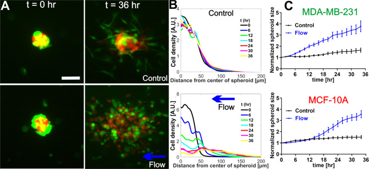
Interstitial flows promoted co-culture tumor spheroid dissociation. (A) Micrographs of co-culture tumor spheroids embedded in collagen matrices of 1.5 mg/mL at t = 0 hour (left panel) and t = 36 hours (right panel) in the absence (top panel) and presence (bottom panel) of the flow. Green: MDA-MB-231 cells expressing EGFP. Red: MCF-10A cells expressing dTomato variants. Scale bar is 100 µm. (B) Quantification of MCF-10A cell distributions using radial cell density in control (top) and flow (bottom). Here, cell density along the y-axis is computed using the azimuthal average fluorescence intensity at a specific radial distance from the center of the spheroid. Each colored line is a radial cell density profile at a specific time point, with t = 0 about 2 hours after the spheroids were introduced into the collagen matrices (C) Normalized spheroid size time evolution for MDA-MB-231 (top) and MCF-10A cells (bottom) with and without flow. Spheroid size is determined by fitting the radial cell density profile to a Gaussian function, and the sigma value is extracted as the spheroid size. Normalized spheroid size is computed as the spheroid size divided by the initial spheroid size at t = 0. We note the initial MDA-MB-231 spheroid size is about 20 percent larger than those of MCF-10A. Interstitial flows significantly increased spheroid size to almost two-fold of its original size. N = 17 spheroids (no flow) and N = 11 spheroids (flow) were used to generate the statistics. Mean and standard error of the mean are shown. ImageJ was used to prepare Fig. 2A.

