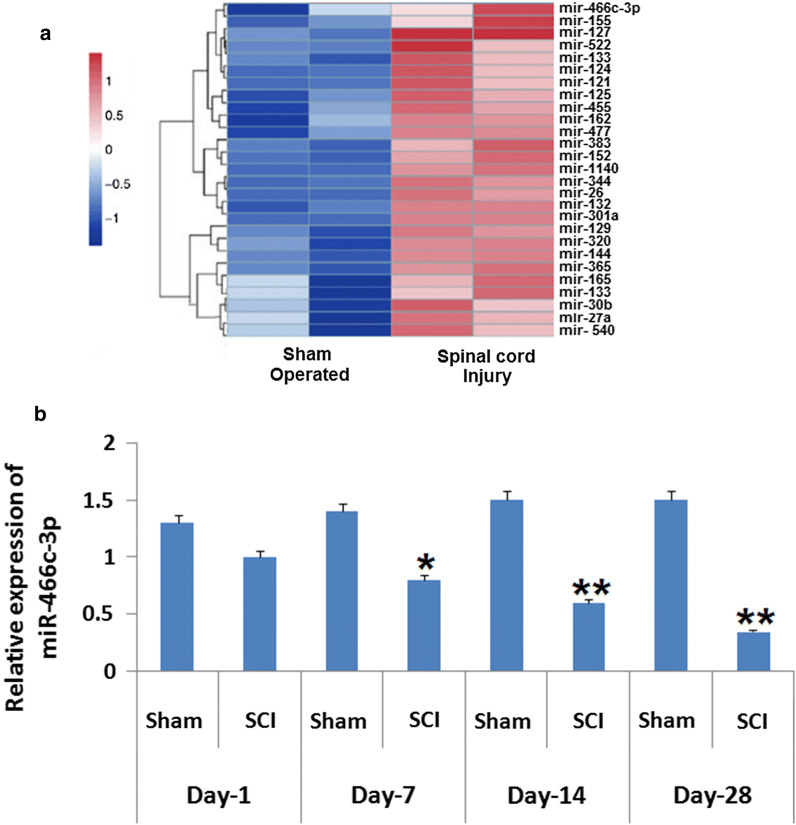
Fig. 1.
Expression of miR in rats subjected to spinal cord injury. a Outcomes of heat map analysis showing significant changes in expression of miRs in rats after 14 days subjecting them to spinal cord injury. The blue color shows suppression and red shows over-expression. b Quantitative results of reverse transcription by qRT-PCR for determining the expression of miR-466c-3p in the spinal cord tissues of rats isolated on the 1st, 7th, 14th and the 28th day after inducing spinal cord injury. The results are presented mean ± SD. *P < 0.05, **P < 0.01 compared to sham operated animals

