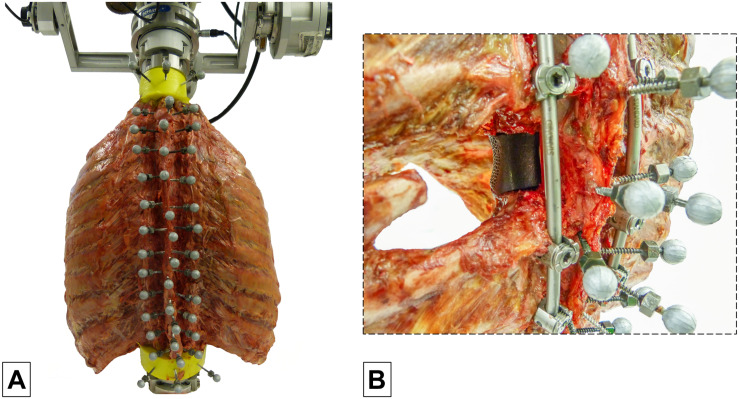
FIGURE 1.
Illustration of the test setup showing a thoracic spine and rib cage specimen with three reflective markers per vertebra (C7-L1) in the spine tester (A) and an example of a specimen with vertebral body replacement implant at T6 level using a unilateral surgical approach stabilized by long posterior instrumentation from T4 to T8 (B).

