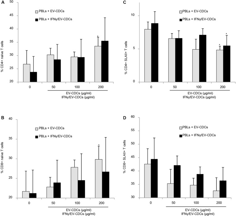
FIGURE 8.
In vitro peripheral blood lymphocyte (PBL) activation and differentiation assays in co-culture with EV-CDCs and IFNγ/EV-CDCs. Peripheral blood lymphocytes (PBLs) were isolated from blood samples by density gradient and co-cultured with 0 (control), 50, 100, and 200 μg/ml of EV-CDC (gray bars) and IFNγ/EV-CDC (black bars) protein for 3 days. Lymphocyte activation/differentiation was analyzed by flow cytometry on CD4+ (A,C) and CD8+ T-cell subpopulations (B,D), using naïve T-cell markers (CD45RA+, CD27+) and activation marker (SLAII). Paired t-test was used to compare doses of EVs to negative controls (*p < 0.05).

