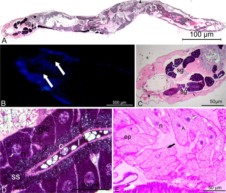
Figure 2.
(A) Longitudinal section of the Orfelia larvae, showing paired tubular structures like the silk glands (sg) from the anterior to the posterior part of the larvae, HE. (B) Total preparation of the cephalic-thorax of Orfelia larvae under fluorescence. Notice blue fluorescence surround the black structures (white arrows). (C) Cephalic-thorax section showing that the black structures saw in B are the silk glands (sg), HE. (D) Detail of the glandular cells (glc) of Orfelia larvae filled with basophil granules (black arrows), which are released to the subcuticular space (ss), crossing the cuticle (c) into the lumen (l). Notice the polytenic chromosomes (white arrow) into the nucleus (n). (E) Histological preparation of the epithelial cells (ep) of Neoditomiya larvae, showing polytenic chromosomes inside the nuclei (n) and vacuolization like (black arrow) into cytoplasm.

