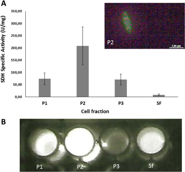
Figure 5.

Cellular location of SBF in Neoditomiya sp larvae: (A) graph of succinate dehydrogenase (SDH) specific activity of cellular fractions. The upper right inset shows the intact mitochondria visualized by fluorescence microscopy; (B) bioluminescent activity of SBF upon mixing extracted cell fractions with Orfelia purified luciferase and 5 mM DTT; (P1) nuclear enriched fraction; (P2) mitochondrial enriched fraction; (P3) peroxisomal enriched fraction and (Sf) final supernatant.
