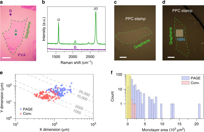
Fig. 2. PVA-assisted graphene exfoliation (PAGE).
Our method allows the use of existing inspection techniques using a optical and b Raman microscopes for identifying monolayer flakes. Scale bar in a is 50 μm. The Raman spectra in the b correspond to the regions in the optical image of the a marked with “A” and “B”. We showed the effectiveness of our layer transfer method by picking up monolayer graphene using c a PPC stamp, and d an hBN/PPC stamp. Scale bars are 20 μm. e PAGE reliably produced very large monolayer graphene flakes, which were not attainable by the conventional method in our experiments. The dashed lines are guides to the eye and represent the product of X and Y dimensions equal to 1000, 2000, 10,000, and 20,000 μm2. f The histogram plot showing the distribution of the monolayer flake area for the samples obtained from 15 consecutive exfoliation trials. The data indicate that PAGE is 20 times more likely than the conventional method (denoted as Conv.) to produce monolayer graphene with an area larger than 1000 μm2 (notice the distribution beyond the yellow shading). The yellow shading denotes flakes with an area of <1000 μm2.

