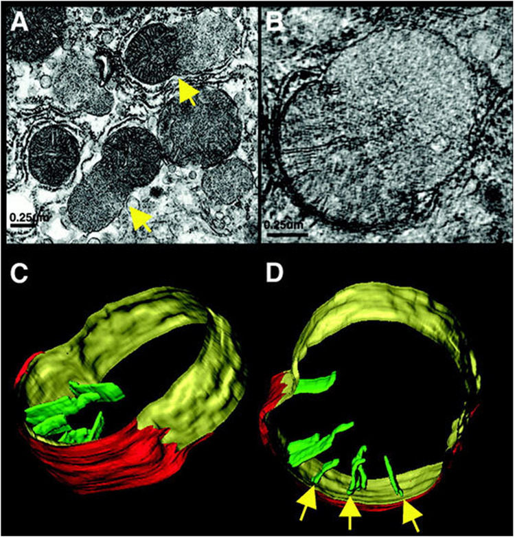
FIGURE 1.
Mitochondrial herniation. (A) Electron micrograph of rat liver, 90 min after FAS activation. Arrow points to a herniation site, a large inner-membrane bleb protruding through a ruptured outer membrane. (B) A slice from an electron tomogram of a herniated mitochondrion. (C,D) Surface-rendered views showing the outer membrane (red), peripheral inner membrane (yellow), and cristae (green). Arrows point to crista junctions. Reproduced from Mootha et al. (2001) with permission (John Wiley and Sons, Inc.).

