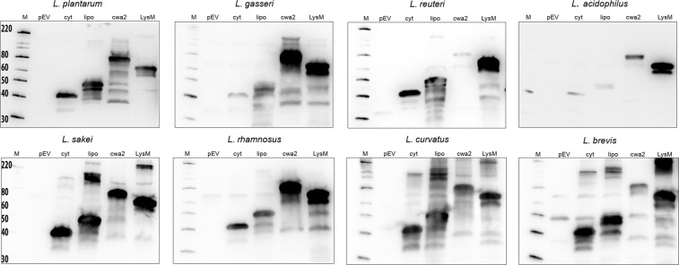
Figure 2.
Production of the antigen. The pictures show western blots of cell-free extracts ofAg85B-ESAT6-DC(AgE6-DC) expressing strains harvested 3 hours after induction. Sample sizes were adjusted to the OD600 of the harvested culture, meaning that all samples represent approximately equal amounts of cells. Lanes: M, molecular mass markers (masses are indicated in kDa); pEV, strain harboring empty vector; cyt, strain harboring vector for intracellular localization (expected mass of the fusion protein is 41 kDa); lipo (48 kDa), cwa2 (69 kDa) and LysM (66 kDa), cell-free extracts of strains harboring various plasmids for anchoring (expected masses between parenthesis). The data presented are from one representative experiment, out of at least three experiments in total. Parts of the lanes marked “lipo” have been published previously30, except in the case of L. sakei.

