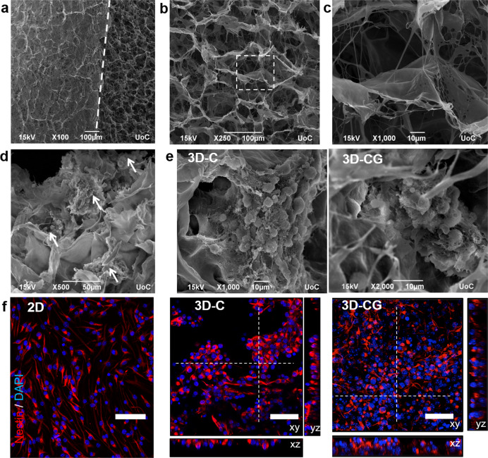
Fig. 1. 3D culture of mouse embryonic neural stem cells inside porous collagen-based scaffolds.
a SEM image highlighting the interface (dashed line) between the scaffold surface (left) and the scaffold interior (right). b SEM image of a porous collagen-GAG scaffold highlighting its porous structure. c High-magnification image of the region shown in b. d SEM image of neurospheres (white arrows) grown inside a collagen-GAG (3D-CG) scaffold at 3 DIV. e SEM images of neurospheres grown inside collagen (3D-C) or collagen-GAG (3D-CG) scaffolds at 3 DIV. f Representative confocal fluorescence images of nestin+ NSCs (red) grown on a PDL-laminin-coated coverslip (2D), inside a collagen scaffold (3D-C) or inside a collagen-GAG scaffold (3D-CG) at 3 DIV. Scale bars, 50 μm.

