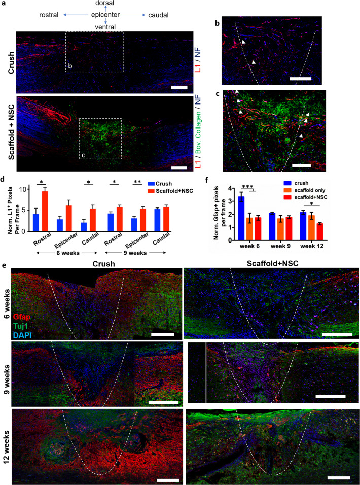
Fig. 5. Porous collagen scaffolds seeded with embryonic NSCs increased axonal elongation and reduced astrogliosis at the lesion site after dorsal column crush.
a Representative fluorescence images of parasagittal sections from crush and scaffold + NSC groups immunostained for neurofilament heavy chain, bovine collagen I and L1, 6 weeks post injury. Scale bars, 200 μm. b, c High-magnification images of regions shown in a highlight axons that stain for NF and L1 at the lesion (inside the graft) or on the approximate lesion boundary (dashed lines). Examples of L1+ axons crossing the approximate lesion boundary are marked by triangles. Scale bars, 100 μm. d Quantification of L1+ pixel density in crush and scaffold + NSC groups, caudally, in the epicenter, and rostrally to the lesion, at 6 and 9 weeks post injury. Results are normalized with respect to the uninjured control group and are presented as mean ± SEM. e Fluorescence imaging of parasagittal sections stained for Tuj1 and GFAP at the lesion 6, 9, and 12 weeks post injury. The approximate lesion boundary is shown using dashed lines. Bars: 200 μm. f Quantification of GFAP+ pixel density in the crush and scaffold + NSC groups at 6, 9, and 12 weeks post injury. Results are normalized with respect to the uninjured control group and are presented as mean ± SEM. “Uninjured control” and “scaffold-only” groups: n = 3. “Crush” and “scaffold + NSC” groups: n = 5. *P < 0.05, ***P < 0.001 (Bonferroni post-hoc pairwise test assuming P1-way-ANOVA < 0.05).

