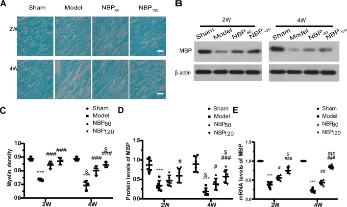
Figure 3.
Dl-3-n-butylphthalide (NBP) attenuates demyelination in corpus callosum after 2VO. (A) Representative images of luxol fast blue (LFB) staining in the corpus callosum at 2 weeks and 4 weeks after 2VO. Bar = 50 μm (n = 3 in each group). (B) Western blot analysis of the expressions of MBP (n = 6 in each group). (C) Myelin density in each group presented by the percentage of stained area in total area of corpus callosum. (D) Quantitative analysis of protein levels of MBP. β-actin was used as an internal control. (E) Quantitative analysis of mRNA levels of MBP. β-actin was used as an internal control (n = 6 in each group). ***p < 0.001, the model group vs. the sham group; #p < 0.05, ##p < 0.01, ###p < 0.001, the NBP60 group or NBP120 group vs. the model group; $p < 0.05, $$$p < 0.001, the NBP60 group vs. NBP120 group; and &p < 0.05, the model group sacrificed at 2 weeks vs. the model group sacrificed at 4 weeks. Values are expressed as mean ± SD. MBP, myelin basic protein.

