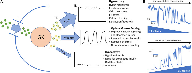
Figure 1.
(A) Glucokinase phosphorylates glucose and feeds the endogenous oscillator. (i). Permissive levels of glucose enter the beta cell through facilitated glucose transport from the blood. (ii). Glucokinase phosphorylates varying amounts of glucose depending on the quantity and activity level of the enzyme along with the blood glucose level. Glucokinase activity levels are largely dependent on the amount of glucose present in the cell but can be augmented in pathological conditions such as hyperinsulinemia and diminished late in the disease process. (iii). High, medium, or low levels of phosphorylated glucose move to the next steps of glycolysis and can activate the endogenous oscillator in the correct range (~5–20 mM glucose in islets from non-diabetic mice). (B) In vitro evidence that GK has high control over islet activity and oscillations. Calcium imaging of islets isolated from CD-1 control mice give proof of concept that reducing glucokinase activity in 20 mM glucose can restore pulsatility. Conversely, stimulating glucokinase activity in 3 mM glucose can generate pulsatility and increase intracellular calcium to the point that no calcium oscillations can occur. This adds support that glucose transporters allow permissive levels of glucose into the cell for glucokinase to be the true glucose sensor. Scale bars represent changes in intracellular calcium (fura-2 am 340/380 nm) as a function of time (min). This figure is based on previously published data in (19).

