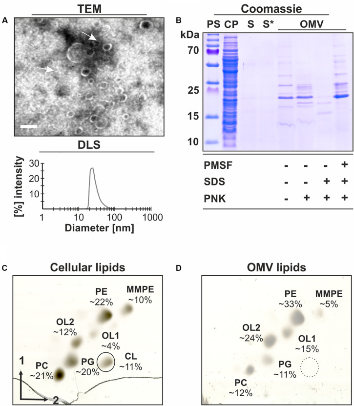
FIGURE 1.
Characterization of OMVs isolated from A. tumefaciens culture supernatants. A. tumefaciens C58 cultures were grown in LB medium to an OD600 value of 1.0 before OMVs were isolated from the cell-free culture supernatants and further characterized. (A) Transmission electron microscopy (TEM) images and size distribution of OMVs determined by dynamic light scattering (DLS). The arrows indicate isolated OMVs. Scale bar: 100 nm. (B) SDS-PAGE analysis and proteinase K (PNK)-protection assays of OMV proteins. (C) Analysis of cellular and (D) OMV lipids by 2-dimensional TLC. Lipids were visualized and quantitated by copper (II) sulfate charring. Lipid standards were used to assign the phospholipid species. PS, protein standard; CP, cell pellet; S, cell-free culture supernatant; S*, S after ultracentrifugation containing secreted soluble proteins; OMV, pellet containing outer membrane vesicles after ultracentrifugation; CL, cardiolipin; PE, phosphatidylethanolamine; MMPE, monomethyl-PE; PC, phosphatidylcholine; PG, phosphatidylglycerol; and OL1/2, ornithine lipids 1/2.

