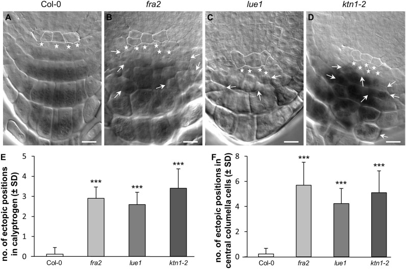
Figure 1.
Ectopic cell divisions in root calyptrogen and root cap malformations. (A–D) Organization of quiescent center and root cap in the root apex of Col-0 (A), fra2 (B), lue1 (C), and ktn1-2 (D) mutants. Cells of the quiescent center are outlined by white dashed line, cells of the columella initials (calyptrogen) are indicated by white stars. Ectopic positions of differentiated cells in the columella are indicated by arrows. (E,F) Quantitative evaluation of ectopicaly positioned cells in the tiers of columella initials—calyptrogen (E) and in columella central cells (F). Data for quantification were collected from 9–17 seedlings. Asterisks indicate statistical significance between measurements (***p < 0.001). Bar: (A–D) 10 μm.

