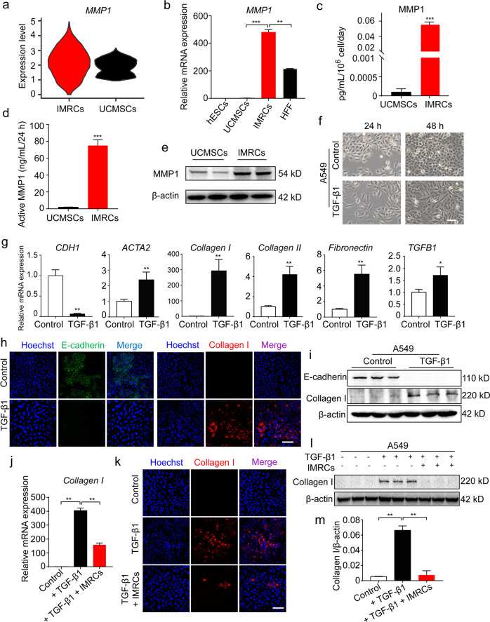
Fig. 4. IMRCs reduce the pro-fibrotic effects of TGF-β1.
a Quantification of MMP1 gene expression amongst single IMRCs, UCMSCs and hESCs, as measured by scRNA-seq. b qPCR for MMP1 mRNA in hESCs, UCMSCs, IMRCs and human foreskin fibroblasts (HFF). c ELISA for MMP1 protein in the conditioned media of IMRCs and UCMSCs. d MMP1 activity in the conditioned media of IMRCs and UCMSCs. e Western blot for MMP1 protein in IMRCs and UCMSCs. β-actin was used as a loading control. f Representative morphology of A549 cells, with or without 10 ng/mL TGF-β1 treatment for 48 h. g qPCR for the relative expression of CDH1, ACTA2, Collagen I, Collagen II, Fibronectin and TGFB1 mRNA in A549 cells, with or without TGF-β1 treatment for 48 h. h Immunofluorescence staining for E-cadherin and Collagen I expression in A549 cells, with or without 10 ng/mL TGF-β1 treatment for 48 h. i Western blot for E-cadherin and Collagen I protein expression in A549 cells, with or without 10 ng/mL TGF-β1 treatment for 48 h. j qPCR for Collagen I mRNA in A549 cells, with or without 10 ng/mL TGF-β1 and IMRC conditioned media treatment for 48 h. k Immunofluorescence staining for E-cadherin and Collagen I expression in A549 cells, with or without 10 ng/mL TGF-β1 and IMRC conditioned media treatment for 48 h. l Western blot for E-cadherin and Collagen I protein expression in A549 cells, with or without 10 ng/mL TGF-β1 and IMRC conditioned media treatment for 48 h. m Quantification of the relative Collagen I protein expression levels in l. *P < 0.05, **P < 0.01, ***P < 0.001; data are represented as the mean ± SEM. Scale bar, 100 µm.

