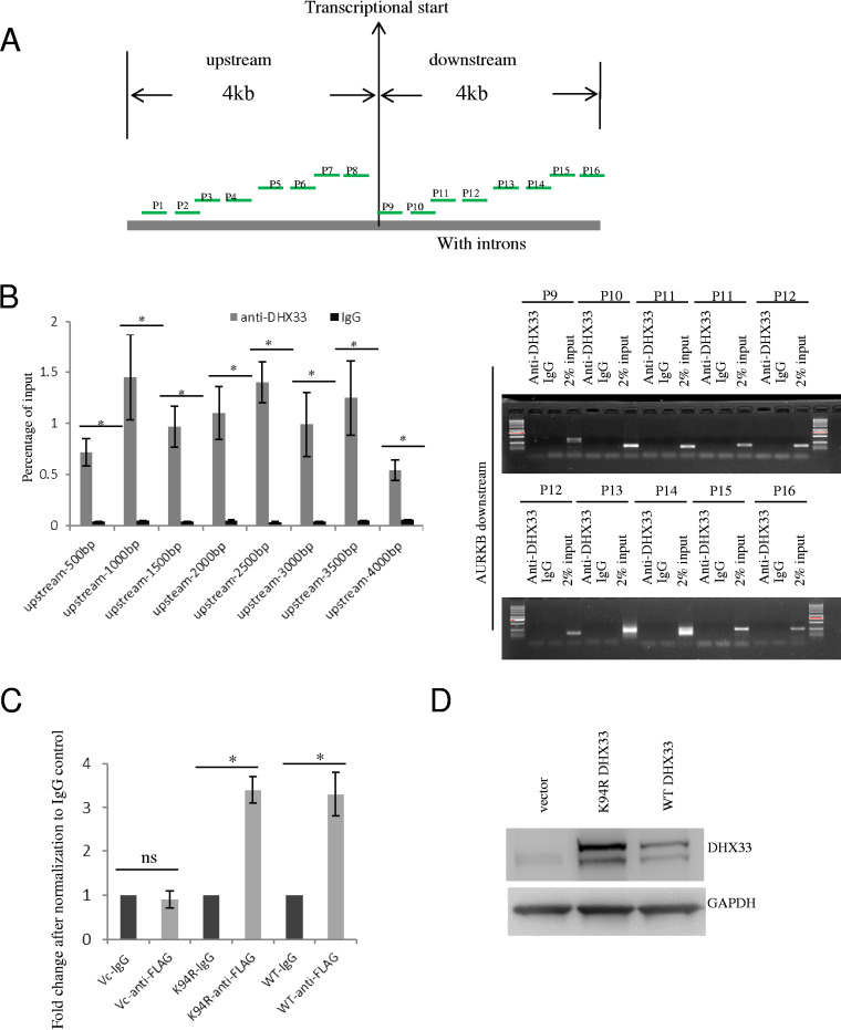
FIG 5.
DHX33 bound to the proximal promoter region of Aurora kinase B gene. (A) Diagram showing the positions of different primer sets for analyzing the binding position of DHX33 on the Aurora kinase B gene. A range of upstream 4 kb to downstream 4 kb of the gene body is chosen. (B) Results of ChIP analysis using the different pairs of primers. H1299 cells were subjected to ChIP analysis with anti-DHX33 antibody; IgG was used as a control. For the upstream gene promoter region, DNA samples were used as described for the quantitative PCR analysis. The data are presented as percentages of input. *, P < 0.05; n = 3. For the downstream 4-kb region of the gene body, data are presented as gel images after PCR amplification; 2% input was used as a control. (C) H1975 or H1299 cells were infected by lentivirus encoding empty vector, K94R mutant of DHX33, or wild-type DHX33. Five days postinfection, cells in equal numbers were subjected to ChIP analysis with anti-FLAG antibody; IgG was used as a control. Primer set 8 was used for PCR amplification. Quantitative PCR was performed, and data are presented as fold enrichment in comparison to the IgG control. *, P < 0.05; n = 3. (D) Western blotting of whole-cell extracts (H1299) with the indicated antibodies.

