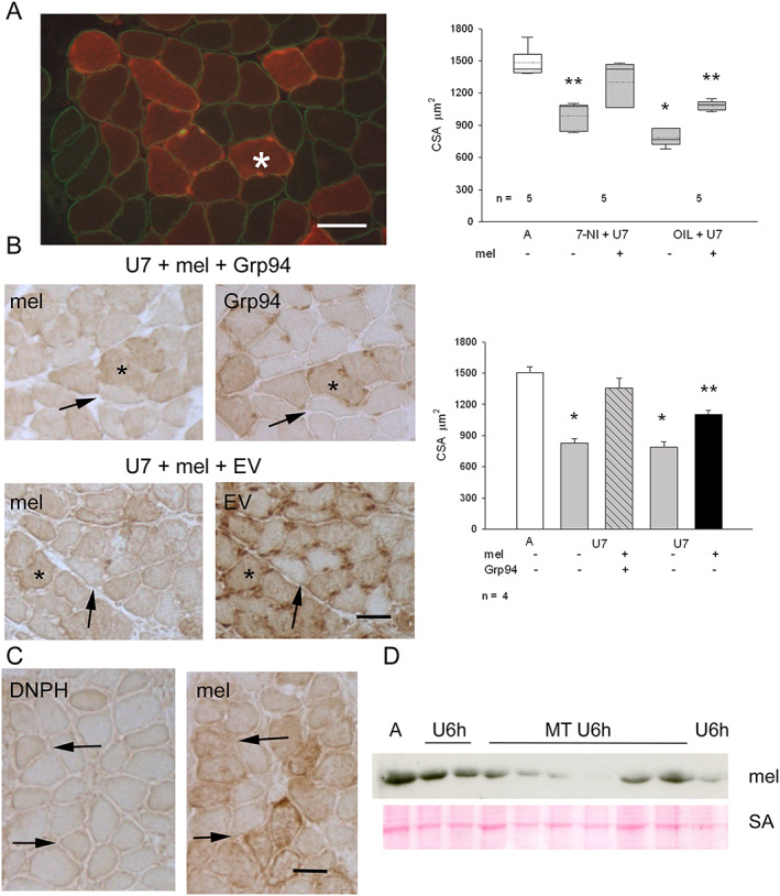
Figure 7.

(A) Left panel: immunofluorescence micrograph from a representative U7 melusin‐transfected soleus from a 7‐nitroindazole (7‐NI)‐treated rat stained for c‐myc tag (red fluorescence), to identify melusin‐transfected myofibers, and dystrophin (green fluorescence) Bar: 50 μm. Right panel displays box plots showing mean (dotted line) and median (solid line) values of cross‐sectional area (CSA) of ambulatory (A) untransfected myofibers measured in EV transfected muscles and 7 day unloaded (U7) melusin‐transfected and untransfected myofibers after treatment with 7‐NI or vehicle (OIL); n indicates the number of muscles studied. A minimum of 100 fibers was considered per group. Asterisks indicate the presence of statistically significant difference vs. CSA values of A and 7‐NI+U7mel+ (P < 0.001, ANOVA and within‐subject ANOVA). Post hoc Tukey's test: P ≤ 0.004 between A and 7‐NI+U7mel−, OIL+U7mel+, OIL+U7mel−; P ≤ 0.05 between 7‐NI+U7mel+ and 7‐NI+U7mel−, OIL+U7mel+ (double asterisk); P < 0.001 between 7‐NI+U7mel+ and OIL+U7mel− (single asterisk); P = 0.14 between A and 7‐NI+U7mel+; post hoc paired Bonferroni's test: P = 0.001 between 7‐NI+U7mel+ and 7‐NI+U7mel− and between OIL+U7mel+ and OIL+U7mel− (double asterisks). (B) Left panels: consecutive cryosections from U7 solei co‐transfected with melusin (mel) and Grp94 cDNA or empty vector (EV) stained in indirect immunoperoxidase with anti‐tag antibodies (c‐myc for melusin and GFP for Grp94 or EV). Asterisk indicates a doubly transfected myofiber; thin arrow an untransfected myofiber. Bar: 50 μm. Right panel: histograms show mean and SEM values of A untransfected myofibers measured in EV transfected muscles and U7 myofiber CSA, measured in doubly transfected myofibers and untransfected ones; n indicates the number of muscles examined. At least 100 myofibers for group were considered for each muscle. Asterisks indicate significant difference vs. CSA values of A and U7mel+Grp94+ (P < 0.001, ANOVA and within‐subject ANOVA). Post hoc Tukey's test: P < 0.01 between A and U7mel−Grp94−, U7mel+EV+ or U7mel−EV−; P ≤ 0.04 between U7mel+Grp94+ and U7mel+EV+ (double asterisk); P ≤ 0.002 between U7mel+Grp94+ and U7mel−Grp94−, or U7mel−EV− (single asterisk); P = 0.22 between A and U7mel+Grp94+; post hoc paired Bonferroni's test: P ≤ 0.005 between U7mel+Grp94+ and U7mel−Grp94− and between U7mel+EV+ and U7mel−EV− (single asterisks). (C) Consecutive cryosections from U7 melusin‐transfected soleus stained in indirect immunoperoxidase with anti‐tag antibodies (anti‐DNPH for carbonylated adducts and anti‐c‐myc for melusin). Thin arrow indicates a carbonylated transfected myofiber; large arrow a carbonylated untransfected one. Bar: 50 μm. (D) Western blot of ambulatory soleus muscles (A) and unloaded ones for 6 h (U6h) in the absence or in the presence of treatment with MitoTEMPO (MT) stained with anti‐melusin antibodies (mel). Red Ponceau staining of serum albumin (SA) is shown as loading reference.
