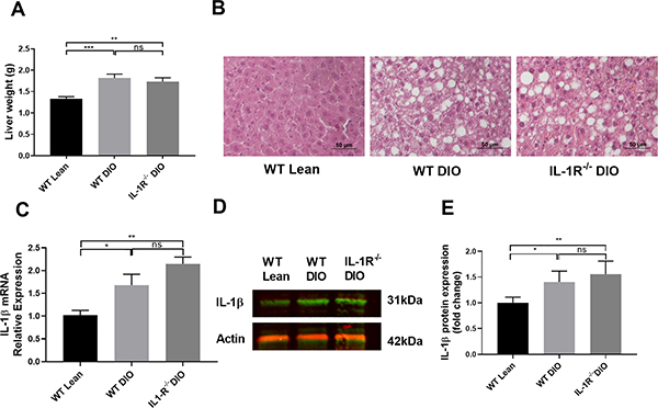
Figure 6. Liver histology and IL-1β expression in WT lean, WT DIO and IL-1R−/− DIO mice.
(A) Liver weight of WT lean, WT DIO and IL-1R−/− DIO mice (n=10 each). (B) HE-stained liver sections from WT lean, WT DIO and IL-1R−/− DIO mice. WT DIO and IL-1R−/− DIO mice showed extensive lipid accumulation compared to WT lean mice. Scale bar: 50μm. (C) Hepatic IL-1β mRNA expression in WT DIO and IL-1R−/− DIO mice (n=5 each). (D) Representative image and (E) Quantification of IL-1β western blot (n=6 each). Data were analyzed using one-way ANOVA followed by Turkey post-test analysis or by Kruskal-Wallis test, followed by Dunn post hoc test. ns=not significant, *p<0.05, **p<0.01, ***p<0.001.

