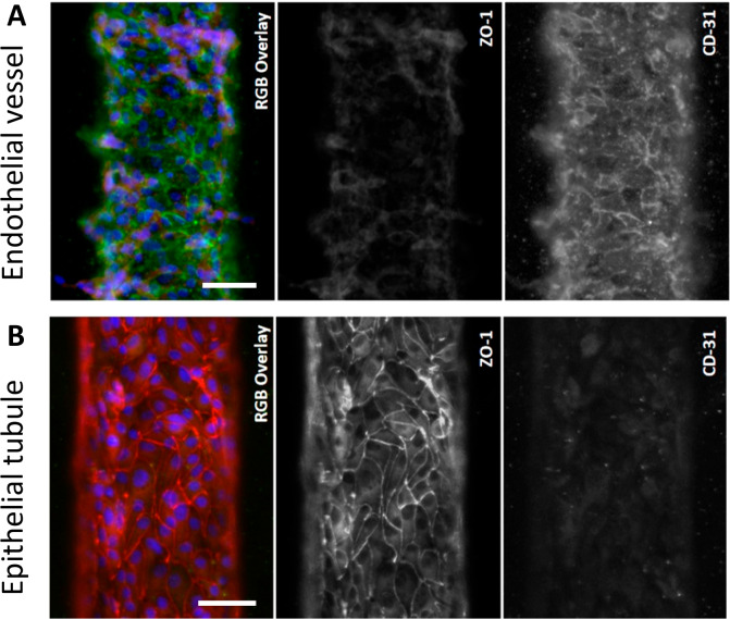
Figure 3.
Morphological characterization of coculture in the VPT-MPS. (A) Endothelial vessel expressing general marker CD-31 (green) but very little ZO-1 (red). (B) Epithelial tubule expressing ZO-1 (red) but very little CD-31 (green). Average epithelial tubule and endothelial vessel diameters are 120 μm. Shown are representative images of cells isolated from a single donor (characterization was reproduced in n = 3 VPT-MPS devices, each from different cell donor). Scale bar: 50 μm.

