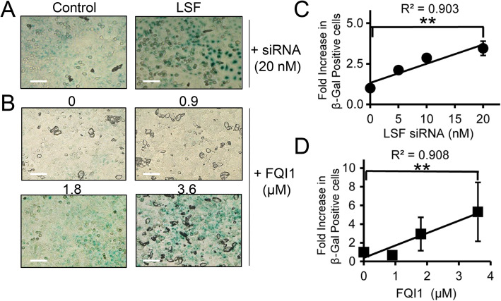
Fig. 6.
Inhibition of LSF activity induces cellular senescence. a-b HeLa cells treated either with increasing concentrations of FQI1 (0 = vehicle control) or with control or LSF siRNA were synchronized using a double thymidine block (protocols in Fig. 2a and 3a, respectively) and then fixed at 8 h after release from the second thymidine block and stained for β-galactosidase activity. Phase contrast images were taken at 20x magnification. Images shown are representative of three independent experiments. c-d The correlation of increasing LSF siRNA concentrations (c) or increasing FQI1 concentrations (d) with the number β-galactosidase positive cells is depicted as a fold change compared to the control for each individual FQI1 and LSF siRNA concentration. The absolute percentage of senescent cells varied somewhat for both controls and experimental samples, ranging for example from 87 to 98% at 20 nM LSF siRNA and 73–96% at 3.6 μM FQI1. The data reflect analysis of 75 cells per condition in each experiment, averaging over three independent experiments. Pearson correlation coefficients are indicated. Scale bars: 50 μm

