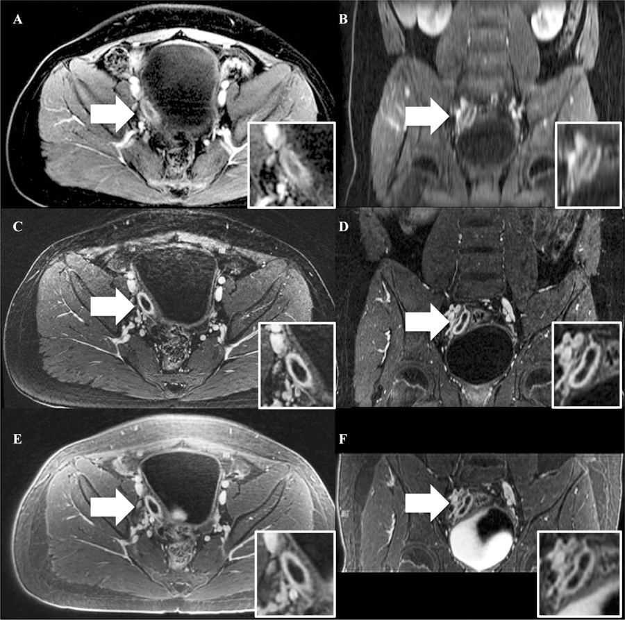
Fig. 3.

A representative case of acute appendicitis labeled on each sequence with white arrows. Note the improved image quality of the coronal reconstructions on FB-SPGR and UTE. A BH-SPGR in the axial plane, B BH-SPGR in the coronal plane, C FB-SPGR in the axial plane, D FB-SPGR in the coronal plane, E UTE in the axial plane, and F UTE in the coronal plane.
