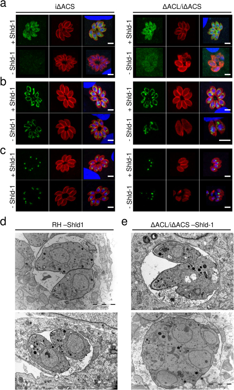
Fig. 2.

Loss of ACS and ACL is associated with amorphic cells and loss of organelle integrity. Immunofluorescence assays (IFAs) of intracellular iΔACS or ΔACL/iΔACS parasites grown in the presence (+) or absence (−) of Shield-1 (Shld-1) for 24 h (a–c). IFAs were fixed and stained with α-gliding-associated protein 45 (α-GAP45, red) to show pellicles of the parasites and 4′,6 diamidin-2-phenylindol (DAPI, blue) to stain the nuclei and either, α-myc (green) to detect ACS (a) or with the monoclonal antibody 5F4 (α-F1B ATPase, green) marking the mitochondrion (b) or α-apicoplast-associated thioredoxin family protein 1 (α-Atrx1, green) staining the apicoplast (c) (scale bars, 5 μm). Electron micrographs of intracellular RH (d) or ΔACL/iΔACS (e) grown in the absence of Shld-1 for 24 h (scale bars, 2 μm). ACL, ATP citrate lyase; ACS, acetyl-CoA synthetase
