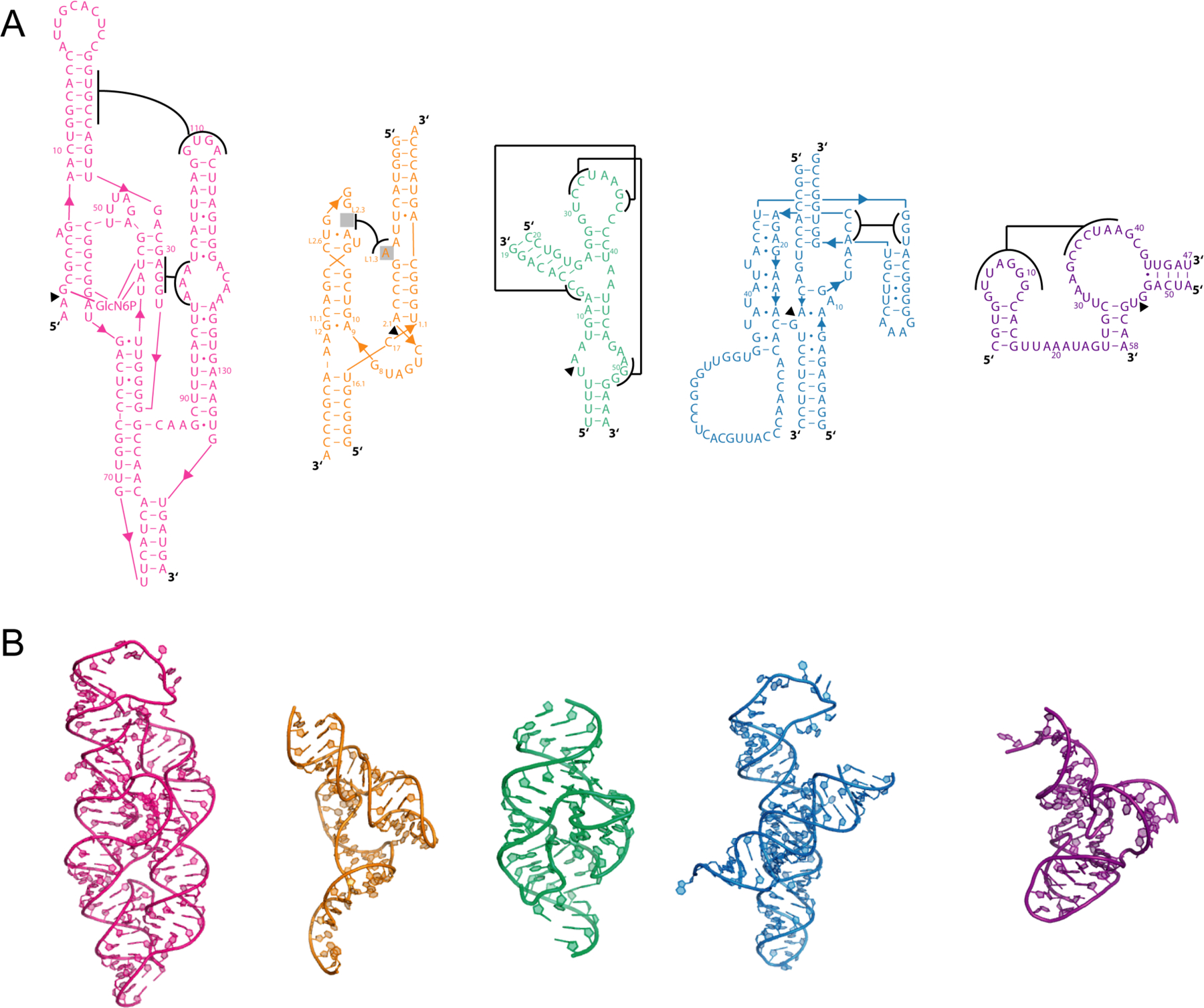
Figure 2.

Structures of ribozymes analyzed in this study. (A) Secondary and (B) tertiary structures for glmS (pink)12, native hammerhead (orange)13, twister (green)14, hairpin (blue)24, and pistol (purple)16 ribozymes. In panel (A), pseudoknot interactions are depicted with brackets adjoined with lines. Tertiary structures were prepared using PyMOL.78
