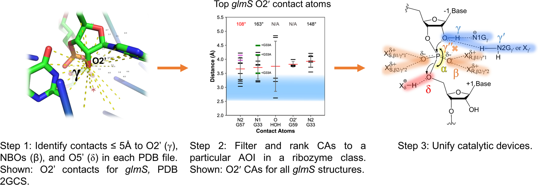
Figure 3.

Automated pipeline to identify ribozyme contacts. Step 1: Contact atoms (CAs) within a specified distance (≤ 5Å) to the atom of interest (AOI), O2′ in this example, were identified and deemed to form a potential contact. Step 2: CAs were filtered according to the occurrence threshold (see Methods) and ranked (L to R) according to their average distance to the AOI; angles for hydrogen bonding are provided above each CA, with optimal hydrogen bonding distance shaded blue. Step 3: Identified contacts were unified to give a mechanistic model depicting strategies for small ribozyme self-cleavage.
