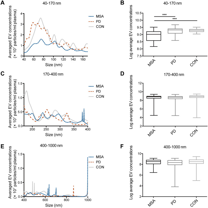
Figure 2.
Fluorescent NTA analysis of plasma CNPase-positive extracellular vesicles in a cohort of patients with MSA, Parkinson’s disease, and healthy controls. CNPase-positive extracellular vesicles in plasma were labelled with anti-CNPase-Qdot605 and analysed directly by using fluorescent NTA for: (A and B) exosome size (40–170 nm, size of exosome plus Qdot) extracellular vesicles, (C and D) 170–400 nm extracellular vesicles, and (E and F) 400–1000 nm extracellular vesicles. The boxes extend from the 25th to 75th percentiles. The middle dark lines indicate the medians. The whiskers extend to the minimum or the maximum values. ***P < 0.001. CON = healthy control; EV = extracellular vesicle; PD = Parkinson disease.

