
Figure 4.
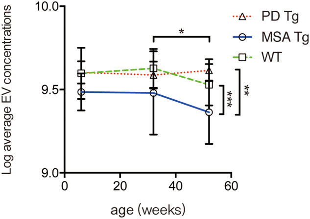
Fluorescent NTA analysis of plasma CNPase-positive extracellular vesicles in a transgenic MSA model, a transgenic Parkinson’s disease model, and wild-type mice. Blood plasma samples were collected from PLP-SYN transgenic (MSA Tg), Prnp-SNCA*A53T transgenic (PD Tg), and wild-type (WT) mice at the ages of 6 weeks, 32 weeks, and 12 months, and the concentrations of CNPase-positive extracellular vesicles were measured using fluorescent NTA. Data shown are mean ± standard deviation (SD). *P < 0.05 (mice at 12 months versus mice at 32 weeks, two-way ANOVA followed by Sidak correction); **P < 0.01 (MSA Tg versus PD Tg, two-way ANOVA); ***P < 0.001 (MSA Tg versus wild-type, two-way ANOVA). EV = extracellular vesicle.