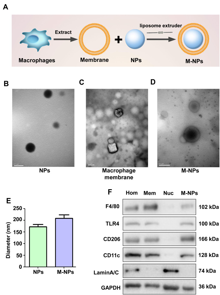
Figure 1.
Formulation and characterization of macrophage membrane-coated nanoparticles (M-NPs). Three-step formulation of M-NPs (A). TEM images of NPs. (Scale bar: 200 nm) (B). TEM images of the macrophage membrane. (Scale bar: 200 nm) (C). TEM images of M-NPs. (Scale bar: 200 nm) (D). The sizes of the NPs and M-NPs were measured by DLS (E). Western blotting was used to detect F4/80, TLR4, CD206, CD11c and lamin A/C protein expression in cell homogenates (Hom), membranes (Mem), nuclei (Nuc) and M-NPs (F).

