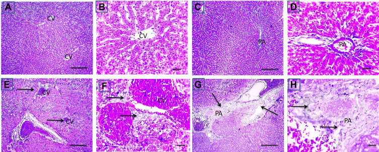
Figure 7.
Effect of CP on fibrosis in CP-induced liver injury. Representative microscopic pictures of Masson trichrome stained liver sections, showing no collagen deposition around central vein (CV) and in portal areas (PA) scored 0 in cont group (A and B) and GL group (C and D). Meanwhile, a moderate stain (E and F) scored 2 to severe (G and H) scored 3 blue stained collagen deposition around central vein (CV) and in portal areas (PA) (black arrows) in group received cisplatin only (E–H). X: 100 bar 100 (A, C, E, and G) and X: 400 bar 50 (B, D, F, and H).

