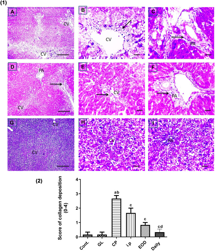
Figure 8.
Effect of GLM on fibrosis in CP-induced liver injury. (1) Representative microscopic pictures of Masson trichrome stained liver sections, showing moderate blue-stained collagen deposition around central vein (CV) and in portal areas (PA) scored 2 (A–C) (black arrows) in i.p group. Mild blue-stained collagen deposition around CV and in PA scored 1 (D–F) (black arrows) in EOD group, no collagen deposition around CV and in PA score 0 (G–I) in daily group. X: 100 bar 100 (A, D, and G) and X: 400 bar 50 (B, C, E, F, H, and I). (2) Statistical analysis of collagen deposition scores in all examined sections of CP-induced liver injury. Collagen deposition scores showing a significant reduction in fibrosis scores in all groups treated with GLM when compared with CP group. Different small alphabetical letters means significant when P< 0.05. aSignificant against control group; bsignificant against GL group; csignificant against CP group; dsignificant against i.p group.

