Figure 12.
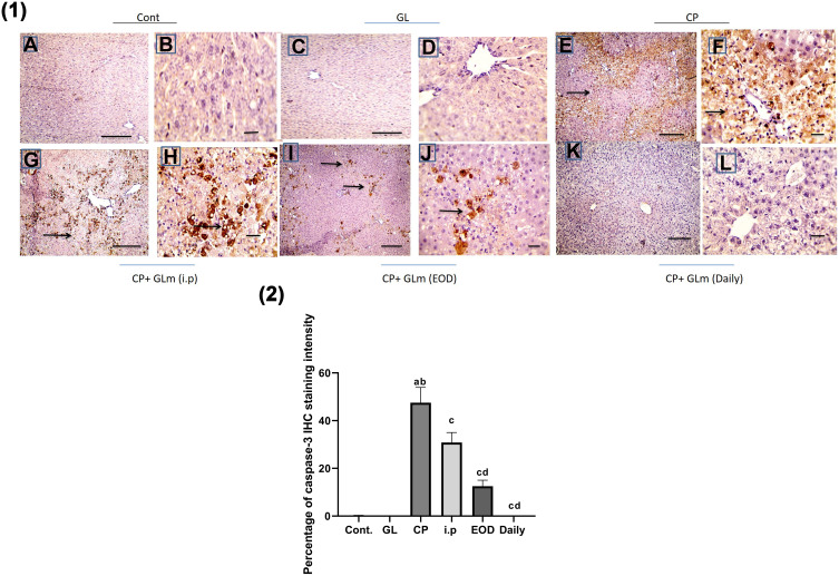
Hepatoprotective effect of Ganoderma lucidum on expression of caspase-3 in rat livers. (1) Representative microscopic pictures of liver sections immunostained using caspase-3 antibody showing negative staining in the control group (A and B) and GL group (C and D), while a strong positive staining as indicated by intense bright brown color in group received cisplatin (E and F). GLM treatment triggers moderate positive staining in i.p group (G and H), mild positive staining in the EOD group (I and J), and negative staining in the daily group (K and L). Black arrows point to positive staining. IHC counterstained with Mayer’s hematoxylin. X: 100 bar 100 (A, C, E, G, and I) and X: 400 bar 50 (B, D, F, H, and J). (2) Statistical analysis of IHC staining intensity percentages in six experimental groups showing a significant increase in caspase-3 in CP group when compared with control and GL groups, and the protective effect of GLM in different treated groups. Different small alphabetical letters means significant when P< 0.05. aSignificant against control group; bsignificant against GL group; csignificant against CP group; dsignificant against i.p. group. (A) Ganoderic acid A binding with HMGB-1 (2D- and 3D-binding modes). (B) Ganoderic acid B binding with HMGB-1 (2D- and 3D-binding modes). (C) Ganoderic acid C6 binding with HMGB-1 (2D- and 3D-binding modes). (D) Ganoderic acid D binding with HMGB-1 (2D- and 3D-binding modes). (E) Ganoderic acid G binding with HMGB-1 (2D- and 3D-binding modes). (F) Ganoderic acid J binding with HMGB-1 (2D- and 3D-binding modes).

