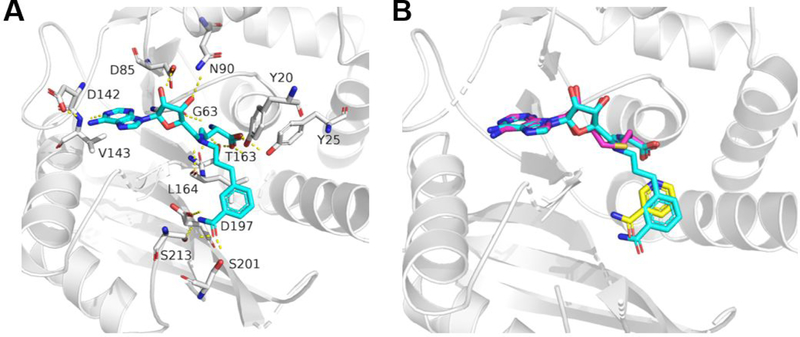
Figure 3.
Docking analysis of compound 1a in the binding sites of hNNMT. (A) Docking of compound 1a (blue) in the hNNMT (gray) structure (PDB 3ROD). H-bond interactions are shown in yellow dotted lines. (B) Overlay of the docking model with the hNNMT (gray)−nicotinamide (yellow)−SAH (pink) complex (PDB 3ROD).

