
FIGURE 3.
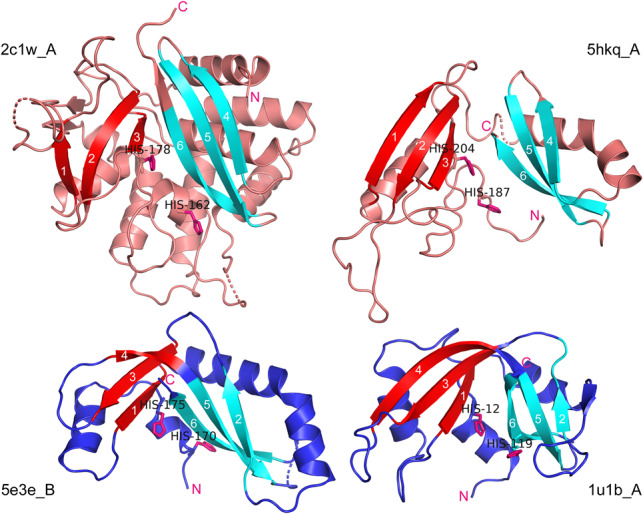
Spatial structure and topology of EndoU, prokRNase A, and animal RNase A proteins. (Upper left) X. laevis EndoU (2c1w_A); (upper right) E.coli EndoU-like toxin (5hkq_A); (lower left) Y. kristensenii RNase A-like toxin (5e3e_B); (lower right) bovine pancreatic ribonuclease (1u1b_A). Known or putative catalytic histidines are shown in all structures. The β-sheets or “wings” of a creased sheet are colored in cyan and red, to match the shading of the strands in Figure 2. The amino-termini in all chains are closely followed by two conserved histidines (or by single His-12 in 1u1b_A), and the carboxy-termini in all chains are located immediately downstream from the conserved Strand 6.