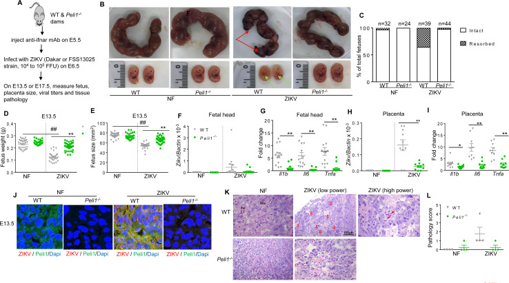
Fig 3. Peli1 mediates inflammatory responses and tissue damage in placenta, and exacerbates birth abnormalities in mice.
WT and Peli1-/- mice were pretreated with MAR1-5A3 at E5.5 followed by infection with 1x 10⁴ FFU ZIKV-Dakar-MA strain one day later. A. Schematic representation of experimental procedure. B. Representative images of E13.5 uteri (upper panel) and fetuses (lower panel). Partial demise and growth restriction was shown in ZIKV-infected WT dams. Red arrows indicate the left placenta residues. Green arrows show growth restriction of fetuses. C. Resorption rates were measured at E13.5. Data are representative of 4–5 independent experiments with one pregnant dam per experiment. D-E. The weight (D) and size (E, CRL x OF diameter) of 24–36 fetuses at E13.5 collected from 4–5 pregnant dams per group. F-I. Viral load (F & H) and cytokine induction (G & I) in fetal heads and placentae collected from 8–14 fetuses per group. D-I: ## P < 0.01 compared to non-infected (NF) group. ** P < 0.01 or *P < 0.05 compared to WT group (Unpaired t test). J. Immunodetection of Peli1 (green), and ZIKV antigen (red) on placentae collected at E13.5. Nuclei are counterstained with DAPI (blue). K-L. Histology staining of placentae at E13.5. K. Representative images (25X) shown are placentae collected from 4 ZIKV-infected dams per group. Scale bar: 100μm (10X). Damaged vessels indicated by hollow arrows; apoptotic bodies indicated by arrows in higher power image; and mild edema indicated by stars. L. Pathology scores were assessed blindly. Data are presented as the means ± SEM.

