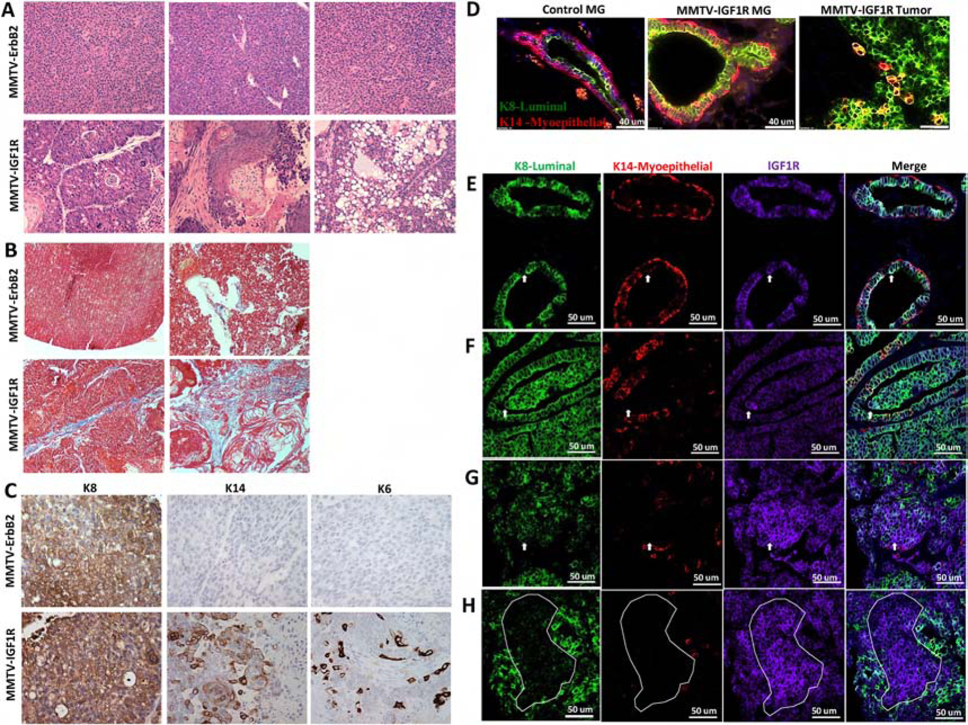
Figure 1: Mammary glands from MMTV-CD8-IGF1R transgenic mice exhibit mixed histologies and multi-lineage tumors.

A) H&E and Masson’s Trichrome staining of MMTV-ErbB2 tumors and MMTV-CD8-IGF1R mammary tumors. 20x magnification. B) Masson’s Trichrome staining of MMTV-ErbB2 tumors and MMTV-CD8-IGF1R mammary tumors. C) IHC of luminal keratin 8 (K8), myoepithelial keratin 14 (K14), and progenitor keratin 6 (K6) markers in MMTV-ErbB2 and MMTV-CD8-IGF1R mammary tumors. Representative images taken at 20x magnification. D) IF of wild type control mammary gland (MG), MMTV-CD8-IGF1R pre-neoplastic mammary gland, and MMTV-CD8-IGF1R mammary tumor co-stained with K8 (green) and K14 (red) (n=5). E) IF of MMTV-CD8-IGF1R mammary gland (n=4) and F-H) IF of MMTV-CD8-IGF1FR tumors (n=5) co-stained with K8 (green), K14 (red), and IGF1R (purple). In (E-F) white arrow indicate example co-stained K8 and IGF1R cell, in (G) white arrow indicates example cell highly expressing IGF1R but with weak K8 expression, and in (H) white dots outline differential staining areas.
