
Figure 3: IGF1R-infected primary mouse MECs demonstrate hyperplastic, highly differentiated outgrowths resulting in attenuated reconstitution.
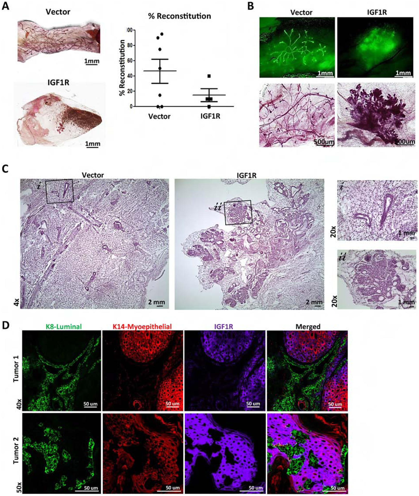
A) Reconstituted glands of wild-type MECs infected with empty vector control (n=7) or CD8-IGF1R lentivirus (n=6). B) Demonstrating ZSGreen and carmine stained mammary outgrowths; 4x and 10x, respectively. C) H&E staining; 4x with 20x insets. D) IF of 15,000 cell reconstituted tumors (n=2) co-stained with K8 (green), K14 (red), and IGF1R (purple).