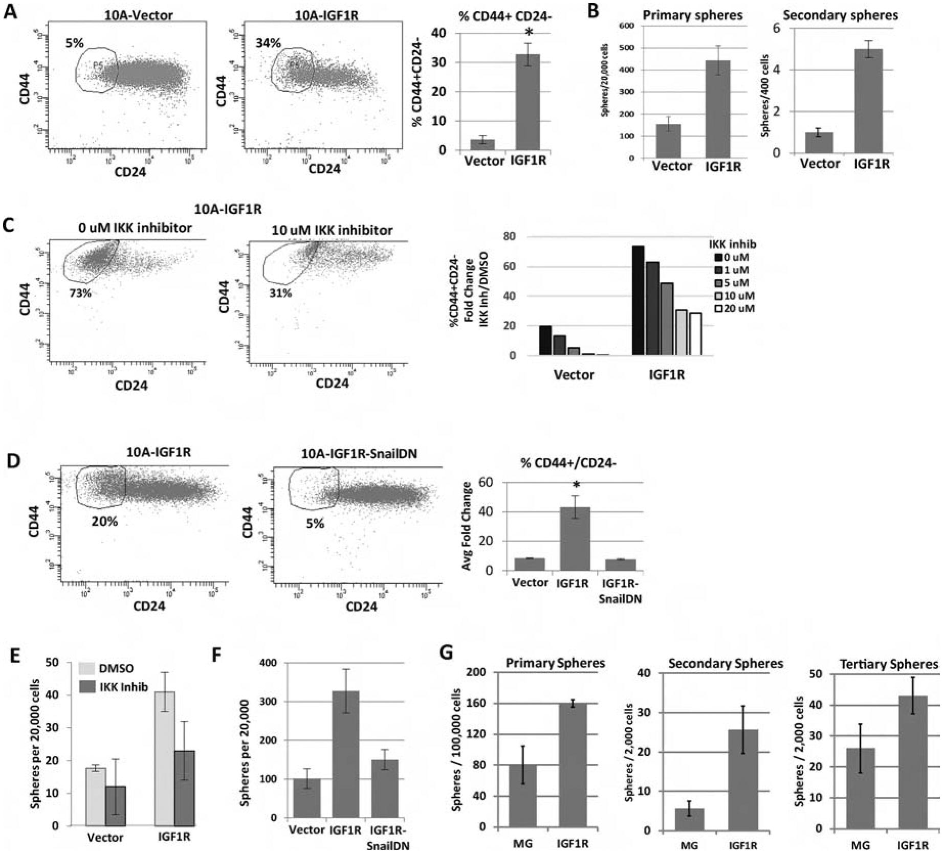
Figure 6: IGF1R constitutive activation expands tumor initiation/stem cell-like characteristics through NFκB signaling and Snail.

A) FACS analysis of the CD44 and CD24 cell surface markers in MCF10A cells overexpressing CD8-IGF1R or empty vector control. Average of 4 experiments with 1 to 2 biological replicates each: t-test p<0.01. B) Sphere formation of MCF10A control and MCF10A-CD8-IGF1R cells grown in serum-free low attachment conditions. 3 independent experiments: t-test p <0.05. C) Representative FACS analysis of CD44 and CD24 with IKK II inhibitor. D) Cells expressing dominant-negative Snail. Quantitative analysis of all inhibitor concentrations is graphed. Representative of 2 (IKK inhibitor) or 3 (SnailDN) experiments. E) Sphere formation of MCF10A-IGF1R cells with IKKII Inhibitor or F) dominant-negative Snail. Representative of 3 independent experiments. G) Sphere formation of MMTV-CD8-IGF1R dissociated tumor cells (see materials and methods). Representative of 3 experiments.
