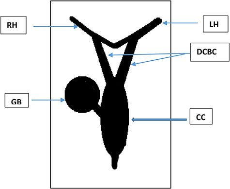
Figure 6.

Schematic illustration of the variant described. LH: left hepatic duct, RH: right hepatic duct, DCBD: double bile duct originating from the hepatic duct confluence, CC: fusiform cyst formed at the distal unified portion of the DCBC, GB: gallbladder emptying into the CC.
