Figure 1.

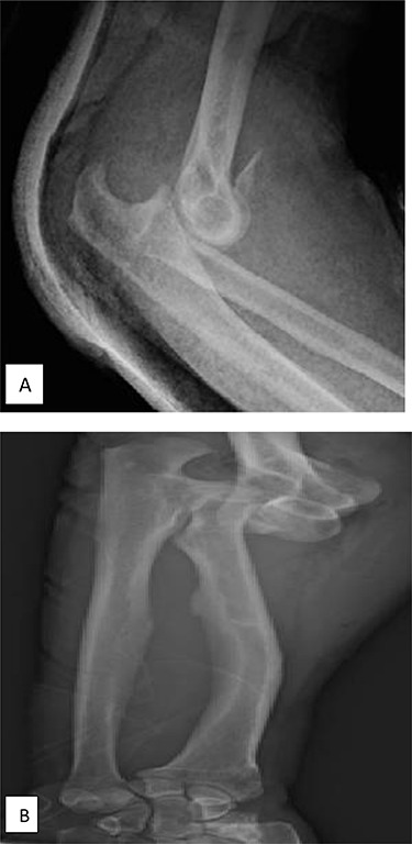
(A) X-ray of elbow profile shows the posterior elbow dislocation with a detached anterior fragment from the coronoid; (B) three-fourth X-ray of the forearm before the reduction demonstrates significant ulnar negative variance.

(A) X-ray of elbow profile shows the posterior elbow dislocation with a detached anterior fragment from the coronoid; (B) three-fourth X-ray of the forearm before the reduction demonstrates significant ulnar negative variance.