
Fig. 1 a.
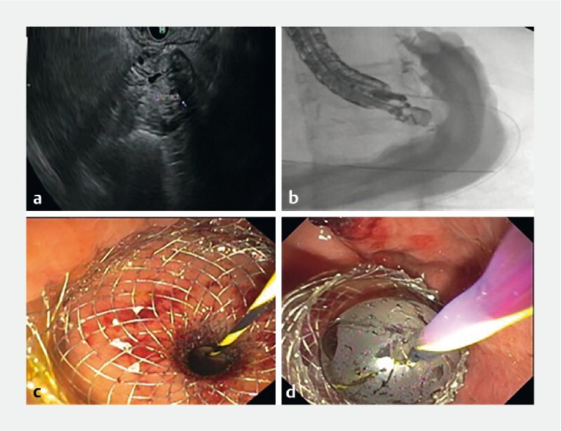
Visualization of excluded stomach through endoscopic ultrasound. b Fluoroscopic imaging showing distension of stomach after injection of contrast. c Deployment of LAMS after creation of a gastro-gastric/gastro-jejunal fistula. d Dilation of LAMS with a CRE Balloon.