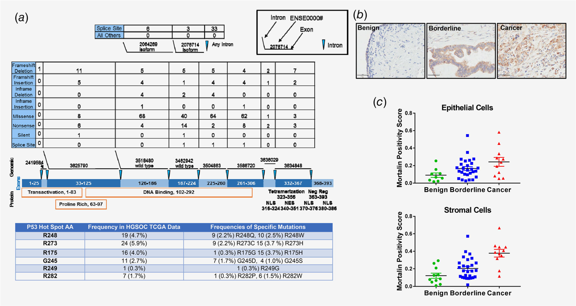
Figure 1.

p53 and mortalin alterations in serous ovarian cancer tissues. (a) Data analysis for detection of p53 mutation status in HGSOC tumors in the TCGA database. The numbers of each type of mutation present are listed above the exons and protein domains depicted. The table shows the most frequently mutated p53 amino acids. (b) Representative images of mortalin immunohistochemical staining from the GOG TMA. (c) Comparison of average mortalin immunohistochemical positivity stain scores for epithelial and stromal cells in benign, borderline and serous tissues. *p ≤ 0.05, ***p ≤ 0.001.
