Figure 6.

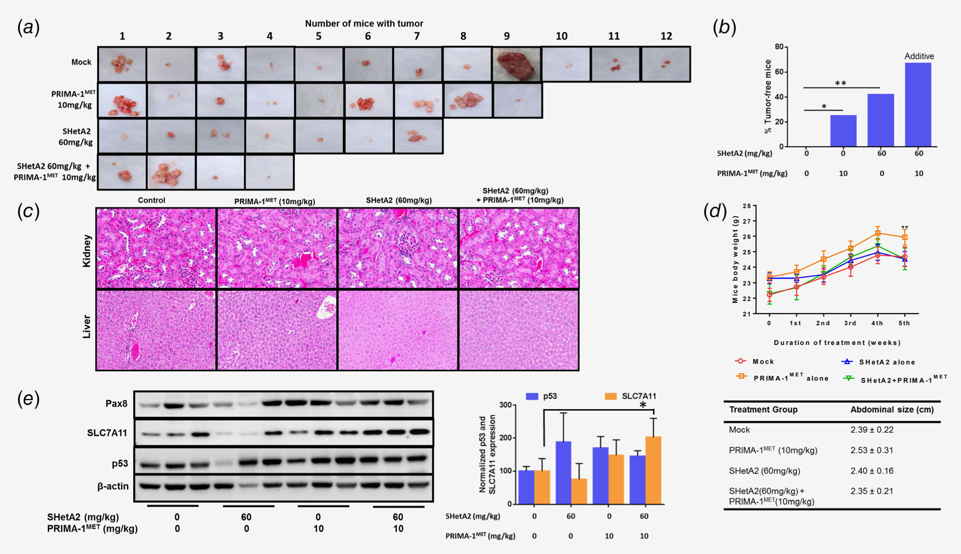
Evaluation of SHetA2 and PRIMA-1MET in model of secondary chemoprevention. (a) Peritoneal tumors harvested from MESOV-injected athymic nude mice after the treatment period. (b) Tumor free rates in the various treatment groups, linear regression model: SHetA2 (p = 0.004, OR = 10.384, 95% CI: 2.158, 48.965) and PRIMA1MET (p = 0.048, OR = 4.464, 95% CI: 1.014, 19.655) functioned additively in preventing tumor development. (c) 20× imaging of H&E staining of liver and kidney specimens of the mouse model to determine the toxicity of drug combination. The kidneys exhibited glomeruli that were well-formed without inflammation and the tubules were normal with no necrosis, apoptosis or inflammation. Livers exhibited no necrosis, fat deposition or inflammation. (d) Mice body weight during the duration of treatment duration and abdominal size observed at the end of the treatment period. ANOVA: ** p < 0.01. (e) Western blot analysis was performed on tumors from three randomly chosen mice in each treatment group to analyze expression of Pax8, SLC7A11 and p53 in mice tumor specimens. Graph shows normalized expression level of p53 and SLC7A11. ANOVA: *p < 0.05.
