
Fig. 4. Ethanol treatment decreases PNPase expression and generates mitochondrial dsRNA in hepatocytes.
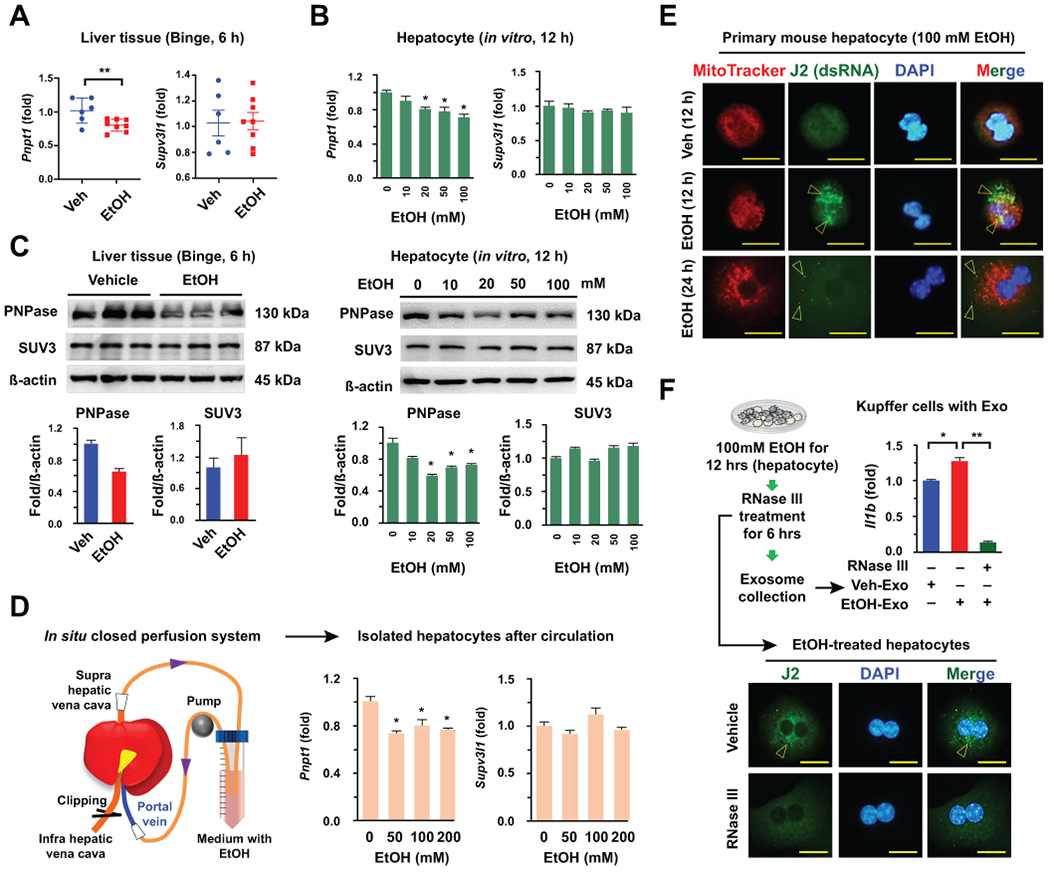
(A-C) Expression levels of genes and proteins were assessed by qRT-PCR and Western blotting in mouse livers (n = 6 ~ 8) and isolated hepatocytes with or without EtOH treatments, respectively. (D) After circulation of EtOH for 2 hours, hepatocytes were isolated and subjected to qRT-PCR. (E) Representative immunostaining of J2 antibody (arrowhead) in Veh- and 100 mM EtOH-treated mouse hepatocyte (12 and 24 hour). Bar = 20 μm. (F) After treatment with 100 mM EtOH for 12 hours, RNase III was added to the medium for an additional 6 hours. Then, hepatocytes were fixed and immunostained with J2 antibody (Bar = 20 μm), and collected exosomes from the medium were co-incubated with freshly isolated mouse Kupffer cells for 24 hours. Kupffer cells were subjected to qRT-PCR. Values and images represent the results from three experimental replicates. Data are expressed as the mean ± SEM. *P < 0.05, **P < 0.01 compared to the corresponding control, based on unpaired t-test between two groups and one way ANOVA with Dunnett’s test for multiple comparison vs control.