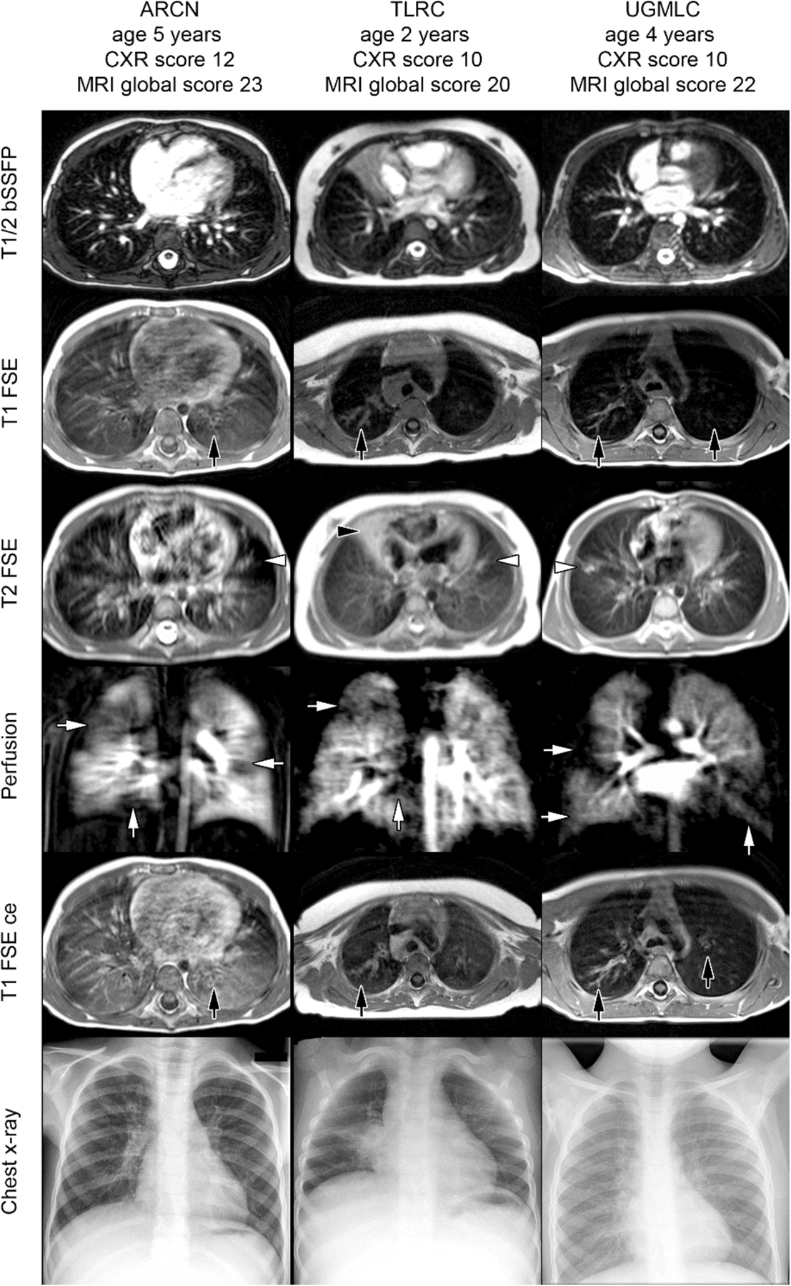
Figure 1:

Examples of T1, T2, and perfusion-weighted images in infants and children with cystic fibrosis. Here, bronchiectasis and wall thickening were identified well on T1-weighted images, with and without contrast (black arrows, T1 FSE and T1 FSE ce, respectively), while mucus plugging and consolidations were identified with T2 weightings (white arrows, T2 FSE). A balanced steady-state free precession sequence (T1/2 bSSFP), contrast perfusion, and chest x-ray are shown for comparison. Reprinted with permission from J Cyst Fibros 2018; 17: 518.
