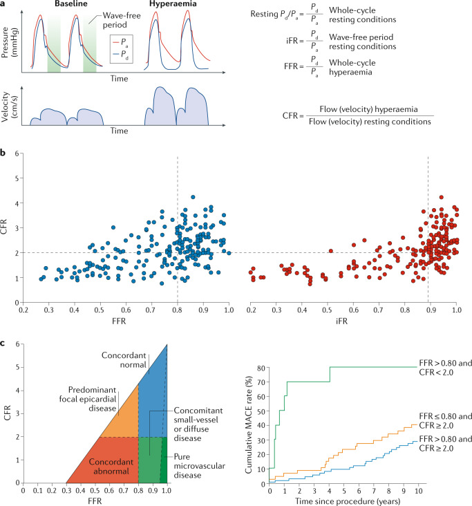
Fig. 8. Invasive coronary flow and pressure measurement.
a | Representation of the ratio of resting distal coronary pressure (Pd) to aortic pressure (Pa), instantaneous wave-free ratio (iFR), fractional flow reserve (FFR) and coronary flow reserve (CFR) calculation from invasively assessed coronary pressure or flow measurement. b | Agreement between coronary pressure (FFR or iFR) and CFR measurement. Discordance between FFR and CFR occurs in 30–40% of individuals, whereas better agreement can be observed between iFR and CFR. c | Interpretation of combined FFR and CFR measurement and its effect on clinical outcome193. Four main quadrants can be identified by the clinical cut-off values for FFR and CFR, indicated by the dashed lines. Patients in the upper right area (blue) have normal FFR and CFR; patients in the lower left area (red) have abnormal FFR and CFR; patients in the upper left area (orange) have abnormal FFR and normal CFR; and patients in the lower right area (green) have normal FFR and abnormal CFR, which indicates predominant microvascular involvement or diffuse coronary artery disease. Patients in the small dark green region in the lower right have an FFR close to 1 and an abnormal CFR, indicating sole involvement of the coronary microvasculature. The prognostic value of FFR and CFR in terms of major adverse cardiovascular events (MACE) is shown on the right. Part c adapted with permission from ref.193, van de Hoef, T. P. et al. Physiological basis and long-term clinical outcome of discordance between fractional flow reserve and coronary flow velocity reserve in coronary stenoses of intermediate severity. Circ. Cardiovasc. Interv. 7(3), 301–311 (https://www.ahajournals.org/journal/circinterventions).

